
Fundus cameras are transforming ophthalmology by enabling faster, accurate, and non-invasive retinal imaging across modern healthcare systems. Today, these advanced tools play a critical role in detecting, diagnosing, and monitoring a wide range of eye diseases effectively. Moreover, retinal imaging devices are widely used to diagnose conditions such as diabetic retinopathy, glaucoma, ocular melanoma, and mild vision issues.
Globally, vision impairment remains a significant challenge, and healthcare systems must adopt advanced diagnostic technologies to address this growing burden efficiently. For instance, approximately 7 million people in the United States experience vision impairment, including nearly 1 million individuals living with blindness today. Therefore, fundus cameras are becoming essential in addressing increasing diagnostic demands and improving clinical outcomes across populations.
Understanding the Role of Fundus Cameras
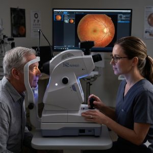
Fundus cameras capture detailed images of the retina, optic disc, macula, and posterior pole with high precision and clarity consistently. Furthermore, these imaging systems allow ophthalmologists to visualize internal eye structures without invasive techniques, improving patient comfort and diagnostic efficiency.
In addition, fundus cameras support early detection of diseases such as diabetic retinopathy, glaucoma, and age-related macular degeneration effectively. Consequently, the healthcare providers can provide the necessary treatment on time to avoid severe problems. Therefore, fundus cameras serve as foundational tools that strengthen clinical decision-making and enhance patient engagement throughout the diagnostic journey.
Growing Demand Driven by Public Health Challenges
The demand for fundus cameras continues to rise due to the increasing prevalence of chronic diseases and aging populations globally. In particular, the United States has a rapidly aging population, leading to more individuals living with diabetes and related complications. Hence, the prevalence of diabetes- and age-related ocular diseases is increasing and needs to be regularly monitored using advanced imaging technologies. Additionally, nearly 2,000 U.S. workers experience work-related eye injuries daily, necessitating immediate medical attention and evaluation.
In addition, the economic burden of major vision problems is expected to rise significantly, reaching an estimated $373 billion by 2050. Thus, there is a need for healthcare systems to be proactive and adopt cost-effective diagnostic tools such as fundus cameras to enable better patient outcomes.
Key Applications of Fundus Cameras in Ophthalmology
Fundus cameras are an indispensable part of modern ophthalmology practices and diagnostic processes, serving a multitude of clinical applications. Firstly, these devices are used to screen for diabetic retinopathy, detect early microvascular abnormalities, and assess retinal damage. Secondly, fundus cameras enable accurate monitoring of glaucoma by capturing high-resolution images of the optic nerve and surrounding structures over time.
Moreover, fundus cameras help diagnose age-related macular degeneration as well as detect retinal tears or detachment during early stages. Hence, these systems are widely used for pre-operative planning and post-operative evaluation, ensuring better clinical outcomes and enhanced patient recovery tracking.
Technological Innovations Transforming Fundus Cameras
Recent technological innovations have greatly improved the efficiency, features, and usability of the fundus camera in healthcare environments. For example, modern fundus cameras use high-resolution sensors and advanced optics to obtain detailed retinal images under challenging conditions. Moreover, portable fundus cameras are revolutionizing point-of-care diagnostics by enabling retinal imaging in remote and underserved locations efficiently.
In addition, artificial intelligence integration has become a key innovation, enabling automated analysis of retinal images for faster and more accurate diagnosis. Furthermore, non-mydriatic fundus cameras eliminate the need for pupil dilation, improving patient comfort while reducing examination time and operational complexity significantly.
The Role of AI and Automation in Fundus Cameras
Artificial intelligence is playing a transformative role in enhancing the diagnostic capabilities of fundus cameras across diverse clinical settings today. AI algorithms can rapidly analyze retinal images and detect early signs of diseases such as diabetic retinopathy and glaucoma accurately. Additionally, automated fundus cameras simplify imaging procedures through features such as autofocus, auto-capture, and guided workflows for consistent image quality. This allows even non-specialist healthcare providers to operate the fundus cameras effectively and to increase access to retinal screening in primary care and community care settings.
Enhancing Accessibility Through Portable Fundus Cameras
Accessibility remains a critical challenge in eye care; however, portable fundus cameras are addressing this issue through innovative design and functionality. For instance, smartphone fundus cameras can be used to take quality pictures of the retina without many infrastructural needs. Similarly, handheld fundus cameras facilitate teleophthalmology, allowing clinicians to transmit retinal images to remote specialists for evaluation. Therefore, patients receive an accurate diagnosis even if they are from isolated geographic regions. Consequently, fundus cameras are helping bridge healthcare gaps and delivering essential eye care services to underserved populations worldwide.
Integration with Telemedicine and Digital Healthcare Systems
Fundus cameras are increasingly integrated into telemedicine ecosystems, improving collaboration, efficiency, and accessibility within healthcare networks significantly. For instance, clinicians can securely share retinal images with specialists for remote consultation and diagnosis seamlessly. Moreover, fundus cameras have been designed to seamlessly interface with digital medical records and cloud systems, making it easy to store and retrieve data for patient management. In addition, automated reporting features reduce administrative workload, allowing healthcare providers to focus more on delivering quality patient care consistently.
Future Trends in Fundus Camera Innovation
The future of fundus cameras is influenced by innovations designed to enhance precision, scalability, and availability in various healthcare systems around the world. One such innovation includes ultra-widefield imaging techniques that allow physicians to visualize a large portion of the retina using a single image. Secondly, advances in computational imaging technologies have enhanced the process of visualization and analysis in retinal imaging, thereby increasing the accuracy of diagnosis for different ocular diseases. Additionally, integration with cloud computing and big data analytics will ensure the effectiveness of population-based screening and predictive diagnostic programs. Efforts to improve affordability and portability will further expand the adoption of fundus cameras in both developed and emerging markets worldwide.
Strategic Outlook
Fundus cameras are revolutionizing retinal imaging by efficiently integrating optics, artificial intelligence, and digital networking in healthcare settings. Consequently, there is an improvement in the early detection and management of eye diseases globally. Further, the issue of public health factors like aging populations and other diseases has made the need for fundus cameras evident in modern healthcare systems. Hence, continuous innovation will keep the fundus camera relevant as a tool in preventing vision loss and enhancing patient outcomes globally.